


tiliGRID-Z
tiliGRID-Z
For businesses, universities and other organizations purchasing for professional use only. No sales to private consumers.
Couldn't load pickup availability


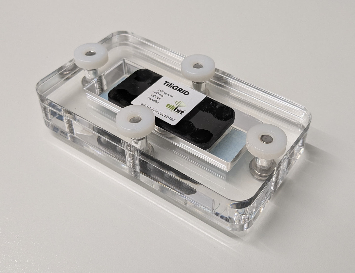
Check three-dimensional localization with a defined DNA reference on a microscopy surface. tiliGRID-Z uses surface-bound L-shaped DNA nanostructures carrying DNA-PAINT docking sites at different heights.
The known geometry supports calibration, comparison of imaging conditions and validation of analysis in localization microscopy. Choose the reference configuration that matches the measurement you need to assess.
Choose your reference
Choose a configuration with 4 or 8 DNA-PAINT handles. Each unit includes an imaging buffer vial and a Cy3b-labeled imager strand. The anti-fouling surface supports low-background single-molecule imaging.
Calibration or sample placement?
tiliGRID-Z provides the imaging reference. To attach your own targets in a local pattern, explore tiliGRID Biotin; for a different geometry or chemistry, discuss tiliGRID Custom.